
Recently, six years of efforts during my Physics Ph.D. have been paid off and the research work about exploring the neuronal activity was published as Wang, Rui, et al. “Ultrasensitive graphene optoelectronic probes for recording electrical activities of individual synapses.” Nano letters (2018). I have to admit that I physically fabricated more than 1000 transistors in the clean room and tested more than 500 microfluidic and graphene devices integrated with neurons. As a Physics graduate, I was able to gain more knowledge in various fields, such as semiconducting engineering, microfluidic mechanics, and biological neuroscience. Furthermore, the collaboration work allows me to grow up as a team leader. Here I want to express thanks to my Ph.D. advisor, Dr. Yaqiong Xu, and other collaborators. This amazing experience paved the way for my research work in brain imaging and my recent transition from a physicist to an engineer.
Abstract about this work:
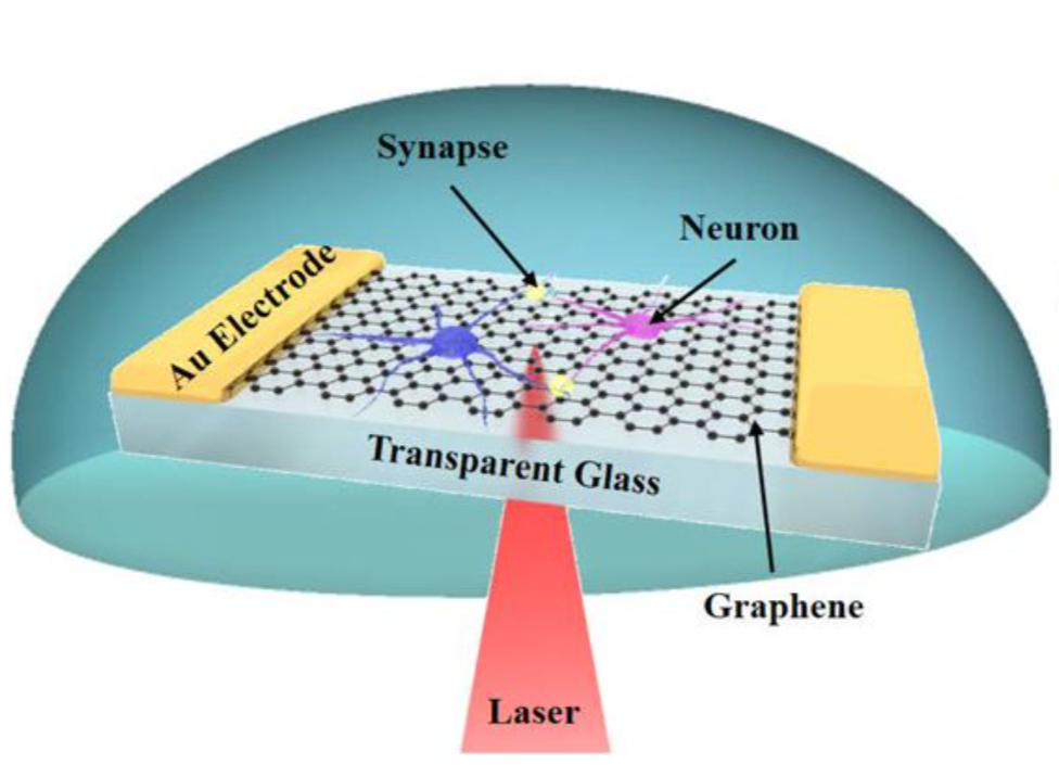
The complex neuronal circuitry connected by submicron synapses in our brain calls for technologies that can map neural networks with ultrahigh spatiotemporal resolution to decipher the underlying mechanisms for multiple aspects of neuroscience. Here we show that, through combining graphene transistor arrays with scanning photocurrent microscopy, we can detect the electrical activities of individual synapses of primary hippocampal neurons. Through measuring the local conductance change of graphene optoelectronic probes directly underneath neuronal processes, we are able to estimate millivolt extracellular potential variations of individual synapses during depolarization. The ultrafast nature of graphene photocurrent response allows for decoding of activity patterns of individual synapses with a sub-millisecond temporal resolution. This new neurotechnology provides promising potentials for recording of electrophysiological outcomes of individual synapses in neural networks.